✍️ Author: Dr Eleni Christoforidou
Home
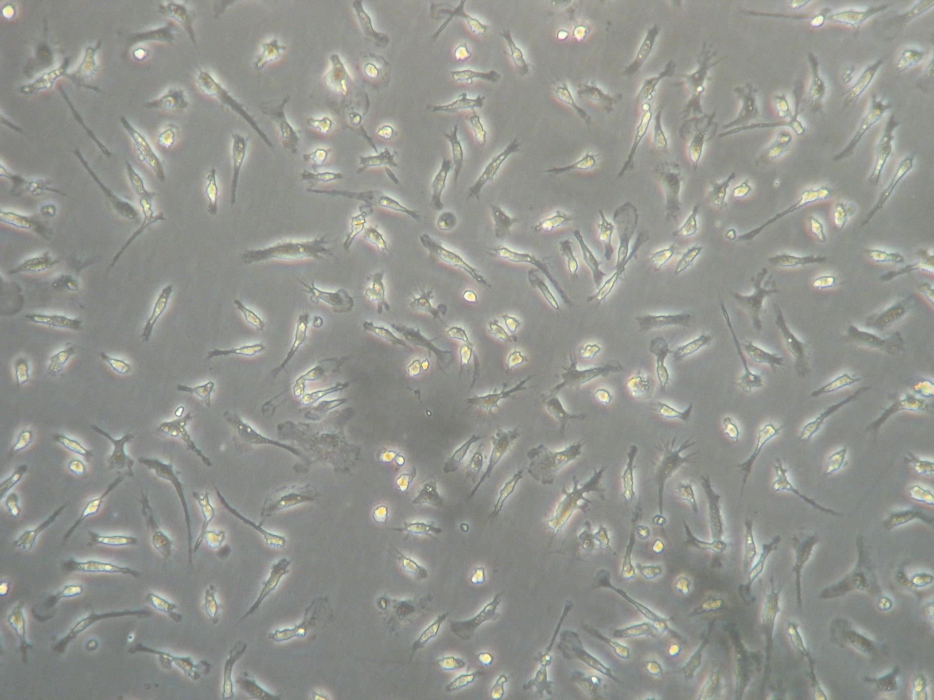
Image from a phase-contrast microscope, capturing microglial cells. As these cells spend more time in culture, they begin to ramify, developing more and more processes.
A Day in the Lab: The Art and Science of Phase-contrast microscopy
🕒 Approximate reading time: 3 minutes
Today, in the lab, my task was twofold: to verify the health of cells thriving in culture and to estimate their population. The confluency of these cells - the degree to which they cover the culture surface - is of prime importance as it informs whether they are ready to take part in upcoming experiments.
One indispensable tool in my daily tissue culture examinations is the phase-contrast microscope. This ingenious instrument allows me to peer into the microscopic universe of these cells, ensuring their growth aligns with expectations.
The beauty of the phase-contrast microscope lies in its capacity to convert subtle phase shifts in light, caused when it traverses through transparent cells, into discernible changes in brightness that construct an image. Although these phase shifts are invisible to the naked eye, they burst into visibility as variations in brightness under the microscope. To achieve this transformation, the microscope expertly separates the illuminating (background) light from the light scattered by the specimen, constituting the foreground details.
A fascinating outcome of this process is that the cells appear to glow - a positive indicator of their health and vitality, as demonstrated in the image below.
Timelapse: Ensuring the robust health and anticipated growth of my glial cells under the watchful gaze of the phase-contrast microscope.